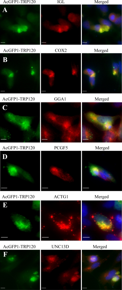
Fig. 5.
Colocalization of IGL, COX2, GGA1, PCGF5, ACTG1, and UNC13D with AcGFP-TRP120 in HeLa cells. pAcGFP1-TRP120-transfected HeLa cells (2 days posttransfection) were labeled and observed by fluorescence microscopy. The AcGFP-TRP120 (green; A to F) and anti-IGL, -COX2, -GGA1, -PCGF5, -ACTG1, and -UNC13D (red; A to F, respectively) signals were merged with 4,6′-diamidino-2-phenylindole staining (blue) (A to F). Bars, 10 μm.

