
Figure 3.
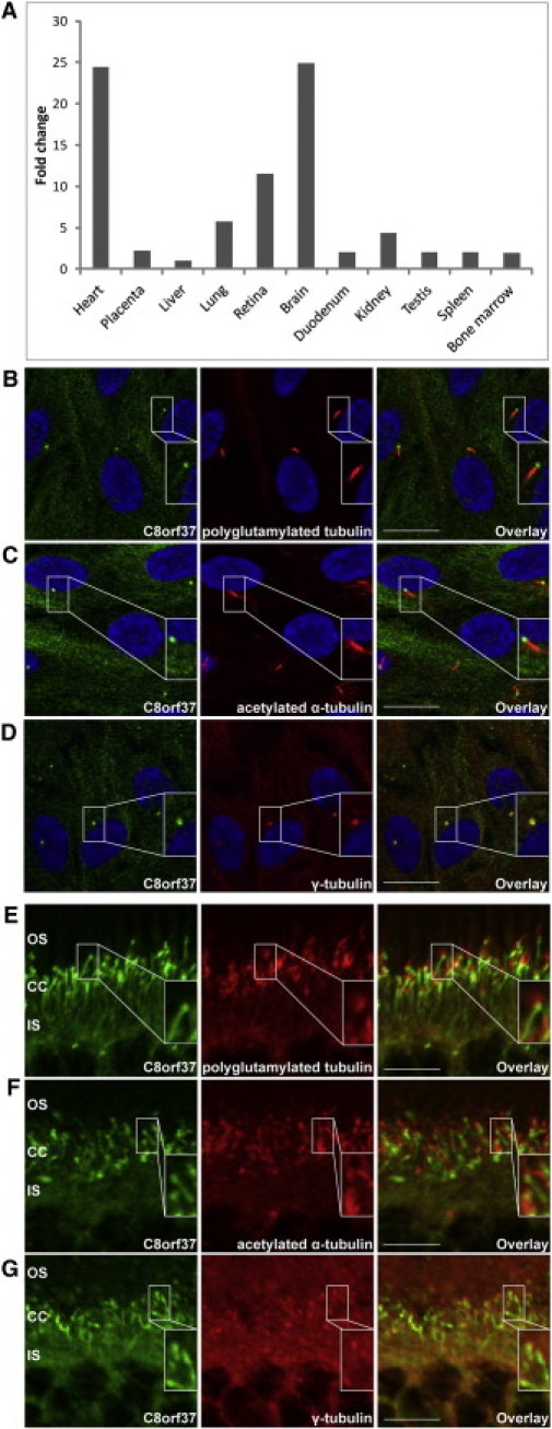
mRNA and Protein-Expression Characteristics of C8orf37
(A) mRNA expression of C8orf37 in adult human tissues is the highest in the heart and the brain, followed by the retinae. The y axis shows the relative expression of C8orf37 in comparison to the lowest expression detected in the liver. For liver expression, the ΔΔCt value was set at 0, resulting in an arbitrary expression of 1.33, 34 The x axis shows the adult human tissues that were tested for C8orf37 expression.
(B–D) Immunohistochemical staining of ciliated hTERT-RPE1 cells with antibodies against human C8orf37 (in green), and (in red) the ciliary markers GT335 (antipolyglutamylated tubulin) (B), antiacetylated α-tubulin (C), and anti-γ-tubulin (D) revealed that endogenous C8orf37 localizes to the base of the primary cilia. Nuclei were stained with DAPI (in blue). Insets show selected magnifications.
(E–G) Immunohistochemical staining in mouse retinal sections (P30). The most intense labeling for C8orf37 (in green) was noted at the base of the photoreceptor connecting cilia which partially colocalized with (in red) the connecting cilium markers GT335 (antipolyglutamylated tubulin) (E), antiacetylated α-tubulin (F), and anti-γ-tubulin (G). C8orf37 also stains structures that extend from the base of the cilium toward the inner segments, suggestive of ciliary rootlets. Abbreviations are as follows: CC, connecting cilia; IS, photoreceptor inner segments; and OS, photoreceptor outer segments. The scale bars represent the following: (B–D), 20 μm and (E–G), 10 μm.