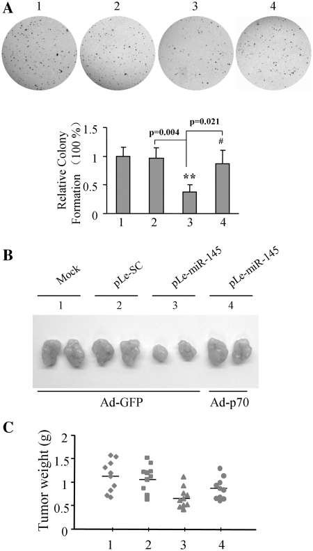
Figure 6.
Role of miR-145 in colony formation and tumor growth. (A) SW1116 mock cells, and cells stably expressing SC and miR-145 were infected with Ad-GFP or Ad-p70S6K1 at 20 MOI for 24 h, then cells were typsinized, and counted. Cells at 5 × 10³ were plated in each well on 6-well plates with 0.05% soft agar layer. After the incubation of the cells for 14 days, the number of cell colonies was counted under the microscope, and the cells were fixed with 100% methanol and stained with 0.5% crystal violet dye. (B) SW1116 mock cells, and cells stably expressing SC and miR-145 were infected with Ad-GFP or Ad-p70S6K1 at 20 MOI for 24 h. Cells were implanted into nude mice. Xenografts were taken out after implantation for 30 days, and represented tumors are photographed. (C) Tumor weight was obtained and presented.

