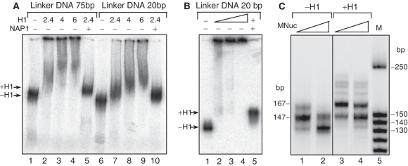
Figure 2.
NAP-1-mediated deposition of the linker histone H1 on dinucleosomes. (A and B) 32P-labelled dinucleosomes with linker DNA of either 75 or 20 bp were incubated with histone H1 alone or with NAP-1-H1 (2:1) complex at the indicated histone H1/dinucleosome ratios. The reaction mixture was then run on native 5% PAGE (A) or 1% agarose gel (B). (C) Miccrococal nuclease digestion of the mononucleosomes. Body-labelled mononucleosomes without H1 (lines 1, 2) and H1-containing (lines 3, 4) were digested with micrococcal nuclease and after arresting the reaction, the digested DNA was purified and run on 10% native PAGE. Lane 5 shows the DNA size marker in base pairs. The positions of the chromatosome band (167 bp) and the core particle (147 bp) are indicated by arrows.

