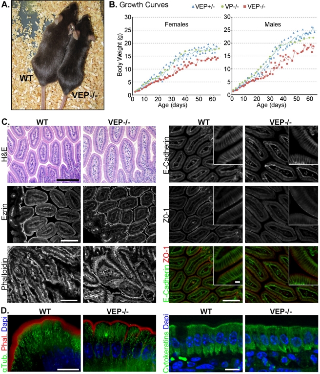
FIGURE 1:
The VEP−/− mice have growth defects, but the morphology of their intestinal epithelium looks normal. (A) Picture of a triple KO VEP−/− animal next to a WT animal of the same age. (B) Growth curves plotting the average weight of 35 triple heterozygotes (VEP+/−), 16 villin/plastin-1 double KO (VP−/−), and 35 triple KO (VEP−/−) mice according to their age. Males and females were analyzed separately. (C and D) Histological sections of jejunum from WT and triple KO (VEP−/−) animals. (C) H&E shows a hematoxilin and eosin staining (scale bar: 200 μm), phalloidin labels F-actin (scale bar: 100 μm). Immunostaining for ezrin, E-cadherin, and ZO-1 is shown. Scale bars: 100 μm; insets: 10 μm. (D) The global cellular organization of the cytoskeletal elements is presented. Microtubules and F-actin are revealed by immunostaining against α-tubulin (α-Tub., green) and phalloidin labeling (Phal., red). Cytokeratins are labeled with a pancytokeratin antibody (green); 4′,6-diamidino-2-phenylindole (DAPI) labels nuclei (blue). Scale bars: 10 μm.

