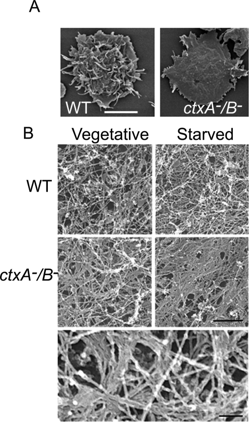
FIGURE 3:
Effects of cortexillin I and II double knockout on cell shape and the actin cytoskeleton. (A) Scanning electron micrographs of vegetative WT and ctxA−/B− cells. Cells were prepared and observed as described in Materials and Methods. Whereas WT cells have a ruffled surface and long filopodia, ctxA−/B− cells have a flat surface and many short spikes at the periphery. Scale bar, 10 μm. (B) Representative transmission electron micrographs showing the actin cytoskeleton organization of vegetative and polarized WT and ctxA−/B− cells that were prepared as described in Materials and Methods. The cytoskeletons of WT cells consist of a largely homogeneous array of actin filaments (top), whereas the cytoskeletons of ctxA−/B− cells contain many bundled actin filament (middle); scale bar, 500 nm. Bottom, enlargement of the vegetative ctxA−/B− cell image showing the F-actin bundles at higher magnification. Scale bar, 100 nm.

