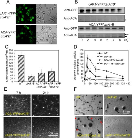
FIGURE 9:
Expression of ACA-YFP in ctxA−/B− cells, but not cAR1-YFP, significantly rescues WT phenotype. About 60% of ctxA−/B− cells expressed cAR1-YFP and ACA-YFP as viewed by confocal microscopy (A). Western blot analysis with anti-GFP antibody confirmed that both proteins were expressed in ctxA−/B− cells (B, top). Expression of ACA-YFP in ctxA−/B− cells increased the expression of endogenous ACA during cAMP pulsing (the solid arrows indicate endogenous ACA, and the dotted arrow indicates ACA-YFP), but expression of cAR1-YFP did not, as detected by anti-ACA antibody (B, bottom). (C) cAMP binding to the cell surface was slightly increased by expression of cAR1-YFP and ACA-YFP in ctxA−/B− cells but was still much less than in WT cells. (D) Adenylyl cyclase A activity was dramatically increased in ACA-YFP/ctxA−/B− cells but not in cAR1-YFP/ctxA−/B− cells. ACA activities were assayed in cells that had been pulsed with cAMP for 7 h, the peak of ACA expression (Figure 5B). (E) ACA-YFP expression in ctxA−/B− cells significantly rescued self-streaming and formation of normal-size mounds but expression of cAR1-YFP did not (see 24-h Supplemental Movies S10 and S11). (F) Development was partially rescued in ACA-YFP/ctxA−B− cells with some complete, although small, fruiting bodies with stem and spore heads (arrows). Development of cAR1-YFP/ctxA−B− cells was not rescued.

