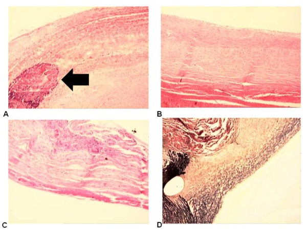
Figure 3.

(A) Aspect of the structure of the patch with calcification (hematoxylin - eosin - 4×). (B) Aspect of the cover layer on the inner face of the patch (RF) (hematoxylin- eosin- 4×). (C) Aspect of the cover layer on the inner face of the patch (SF) (hematoxylin - eosin - 4×). (D) Aspect of the cover layer on the inner face of the patch (RF) (Verhoeff - 4×). Arrows indicate calcification.
