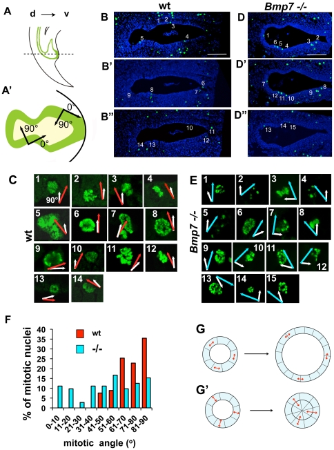
Figure 6. Polarity of cell divisions is disrupted in Bmp7 null the cloacal endoderm.
Sagittal (A) and transverse (A′) views of murine cloacal region at E11.5. Dashed line in (A) indicates the plane of sections in (B–B″, D–D″). Dorsal, d, and ventral, v. (A′) Radial, apical-basal, direction in the cloacal cavity is defined as 90 degrees, and tangential direction, as zero degrees. Examples of transverse cloacal sections at E11.5 of wild-type (B–B″) and Bmp7 null (D–D″) embryos labeled for pHH3 and imaged using confocal microscopy. Scale bars, 100 µm. (D, F) Images of individual wild type (D) and Bmp7 null (F) mitotic nuclear pairs numbered in C–C″ and E–E″ shown at 63× resolution. White vectors indicate the radial direction. Red vectors in (D) and blue vectors in (F) indicate the direction of separation of the chromosome bundles determined on confocal z-sections. (G) Graphic presentation of the distributions of mitotic angles in wild type (red bars) and Bmp7 null (blue bars) cloacal endoderm. KS test: Nwt = 172, Nmut = 168, P<0.001. (G, G′) A simple model of the axial longitudinal section of the cloaca is depicted as a circle, one cell wide and eight cells in diameter. (G) If cell divisions are oriented tangentially (double arrows), after 8 divisions the width of the epithelium remains the same and the diameter of the lumen is increased by 1.7. (G′) If cell divisions are oriented radially, after 8 divisions, daughter cells fill the section and topologically separate dorsal and ventral lumens.

