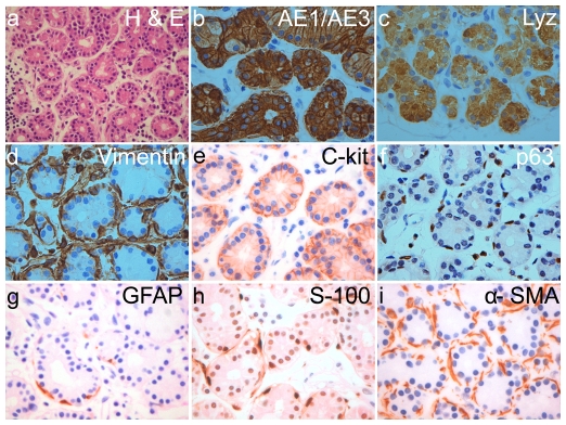
Figure 3. Immunohistochemistry on normal human lacrimal gland.
H&E staining shows the normal histology of the lacrimal gland. Marker staining pattern shows localization of pan-cytokeratin (AE1/AE3) and lysozyme (Lzy) in the cytoplasm of the acinar cells while c-kit is seen in the plasma membrane of acinar cells. p63, glial fibrillary acidic protein (GFAP), S-100 protein and ∝-SMA localize in the myoepithelial cells enveloping the acinar cells. Vimentin is seen in the myoepithelial cells and also in some of the acinar cells. All images are at 40× magnification except H&E which is at 10×.

