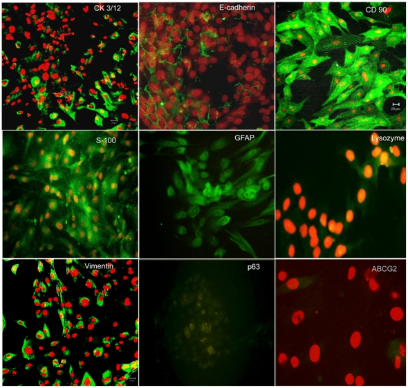
Figure 4. Immunocytochemistry on in-vitro cultured human lacrimal gland cells.
Cells with epithelial morphology stain positively with E-cadherin, CK3/12, lysozyme and p63; oval and plump cells stain positive for myoepithelial markers GFAP and S100 protein while the spindle shaped cells are seen to be positive for mesenchymal markers CD90 and vimentin. Some cells also show immunopositivity for ABCG2. Secondary antibody uses is fluoresceine isothiocyanate (green) and the counter-stain is propidium iodide (red).

