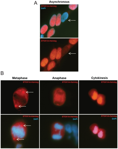
Figure 1. Expression analysis of STOX1A in stably transfected SH-SY5Y cells.
(A) Immunofluorescence shows exclusively nuclear or cytoplasmic (white arrows) staining for STOX1A-Halotag protein in STOX1A stably transfected SH-SY5Y cells. (B) Cells undergoing mitosis showing a non-overlapping STOX1A/DAPI immunofluorescence pattern during metaphase and anaphase until cytokinesis when STOX1A immunofluoresence overlaps with DAPI (DNA) immunofluorescence.

