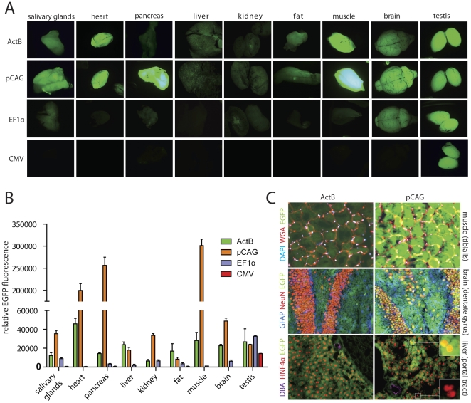
Figure 2. Comparison of different ubiquitous promoters in the modRosa26LoxP locus.
(A) Organs from transgenic mice showing different intensity of overall EGFP fluorescence under low-magnification microscopy, using the same conditions. ActB mice were used as controls for high EGFP expression levels. The pCAG promoter shows the highest overall fluorescence. The CMV promoter showed strong activity only in testis. (B) EGFP fluorescence was quantified in organ homogenates from mR26-pCAG-EGFP, mR26-EF1α-EGFP, mR26-CMV-EGFP and ActB mice. In most organs, EGFP fluorescence was highest in mR26-pCAG-EGFP mice, except fat tissue where ActB mice showed higher levels. Heart, pancreas and muscle showed extremely high levels of EGFP fluorescence. Background fluorescence determined in organ homogenates from wt mice was subtracted (n≥3 mice per genotype). Values are shown as mean ± SEM. (C) Exemplary pictures from cryosections showing strong and ubiquitous EGFP expression in muscle (costained with wheat germ agglutinin (WGA) for muscle fiber wall and DAPI) and brain (hippocampus, costained with NeuN for neurons and GFAP for astrocytes). Mosaic EGFP fluorescence was detected in livers of mR26-pCAG-EGFP mice (costained with HNF4α for hepatocytes and DBA for bile ducts). Magnified insets show both EGFP+ and EGFP- hepatocytes.

