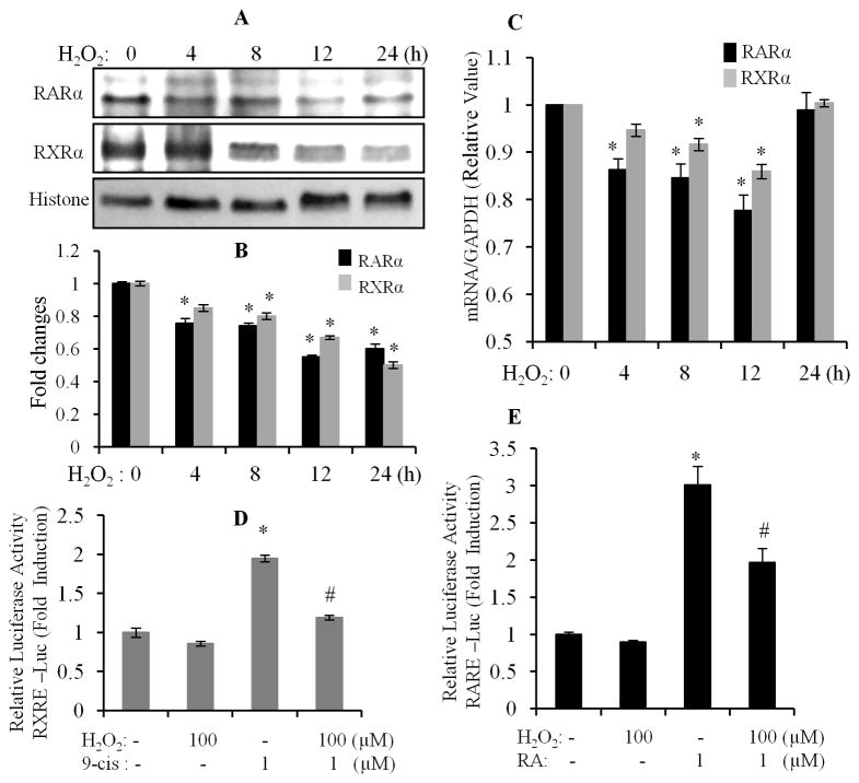
Fig. 4. H2O2 inhibits gene and protein expression of RARα and RXRα.
Cardiomyocytes were exposed to 100 μM of H2O2 for different time periods, and nuclear protein (A & B) and gene (C) expression of RARα and RXRα was determined. Each value represents the mean ± SEM (n = 3). *, p<0.05, versus control. D & E. Cardiomyocytes were transfected with pRXR-Luc and pRAR-Luc, for 6 h, and exposed to 1 μM of 9-cis RA (9-cis) and ATRA (RA), in the absence or presence of 100 μM of H2O2, for 12 h. RARE and RXRE-dependent luciferase activity was measured, as described in Fig. 1. Each value represents the mean ± SEM (n = 3). *, p<0.05, versus control; #, p<0.05, versus HG.

