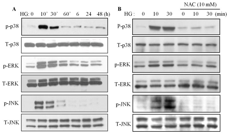
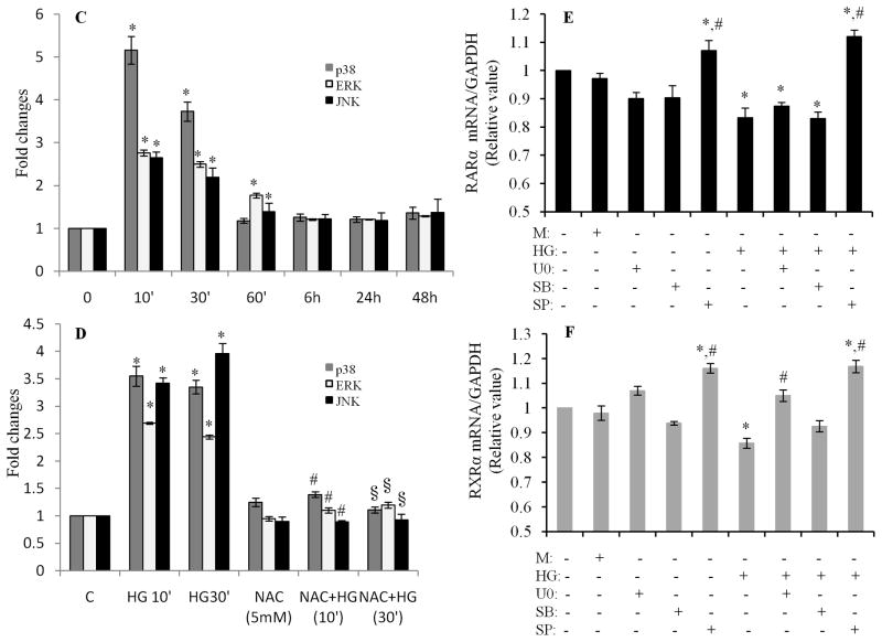
Fig. 5. Role of MAP kinases in regulation of the HG action on RARα and RXRα.
A. High glucose induces phosphorylation of MAP kinases. Cardiomyocytes were exposed to HG for different time periods, and the phosphorylation of p38, ERK1/2 and JNK was determined by Western blotting. C. Intensity of the phosphorylation of p38, ERK and JNK (A) was analyzed by densitometry and standardized to the corresponding total proteins. Data are expressed as the mean ± SEM (n=3). *, p<0.05, versus non-treated group. B. Oxidative stress is involved in HG-induced phosphorylation of MAP kinases. Cardiomyocytes were pretreated with NAC for 30 min and exposed to HG for 10 and 30 min, and the phosphorylation of MAP kinases was determined and quantified (D) as noted in C. *, p<0.05, versus non-treated group; #, p<0.05, versus HG 10 min; §, p<0.05, versus HG 30 min. E & F. Role of MAP kinases in regulation of HG-induced downregulation of RARα and RXRα. Cardiomyocytes were pretreated with 10 μM of U0126, SB203580 or SP600125 for 30 min, and exposed to HG for 12 h. Gene expression of RARα (E) and RXRα (F) was determined. Data (mean ± SEM, n = 3) are expressed as a relative value compared to control. *, p<0.05, versus control; #, p<0.05, versus HG.


