
Fig. 2.
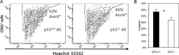
Ex-vivo cultured p53−/− Mk cells exhibit diminished apoptosis compared to p53+/+ Mk cells on day 4 of Tpo culture. Cells were incubated with 0.01 mM Hoechst 33342, which serves to distinguish nucleated Mk cells from platelets and other CD41+ debris in the culture, stained with anti-CD41 and treated with Annexin V to identify apoptotic Mk cells. (A) Representative apoptosis data from p53+/+ and p53−/− cultured Mk cells and (B) average of the apoptosis data for day 4 of culture. Error bars: SEM, N=3, * indicates P < 0.05.