Fig. 5.

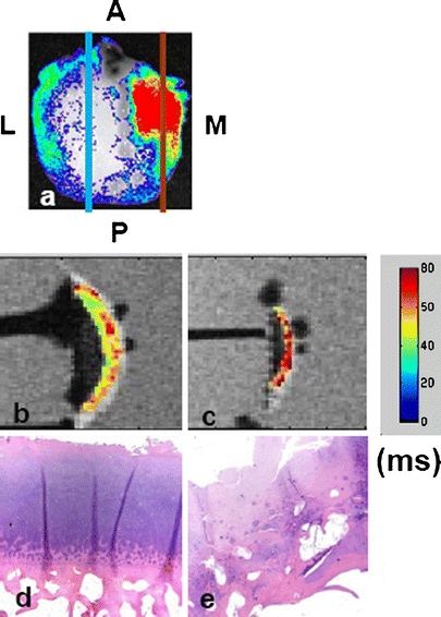
A femoral condyle (K040MPFC) was treated with MMPSense 680 and examined by optical and MR imaging. The optical signal was normalized against the background excitation and expressed in photon efficiency. The specimen was imaged in its respective physiological positions to simulate the in vivo orientation and position: M medial, L lateral, A anterior, and P posterior. Signals observed here indicate high MMP activities in the medial compartment (a). Two slices were removed from high (red) and low (blue) optical regions for histological analysis. b, c Color-coded T 1ρ maps overlaid on a 3-Tesla MR image with (b) corresponding to the low optical signal region and (c) corresponding to the high optical signal region. The T 1ρ relaxation value is ranging from 40 to 60 ms with the highest value coded as red. The intense T 1ρ region signifies the severe loss of proteoglycans. d, e The hematoxylin and eosin staining showing the region with low MMP optical signal (d) has minimal clefting, normal tidemark, and normal cellularity. On the contrary, the region with high MMP optical signal exhibits severe fissuring and clefting, disorganized tidemark as well as the loss of cellularity.
