Figure 1.
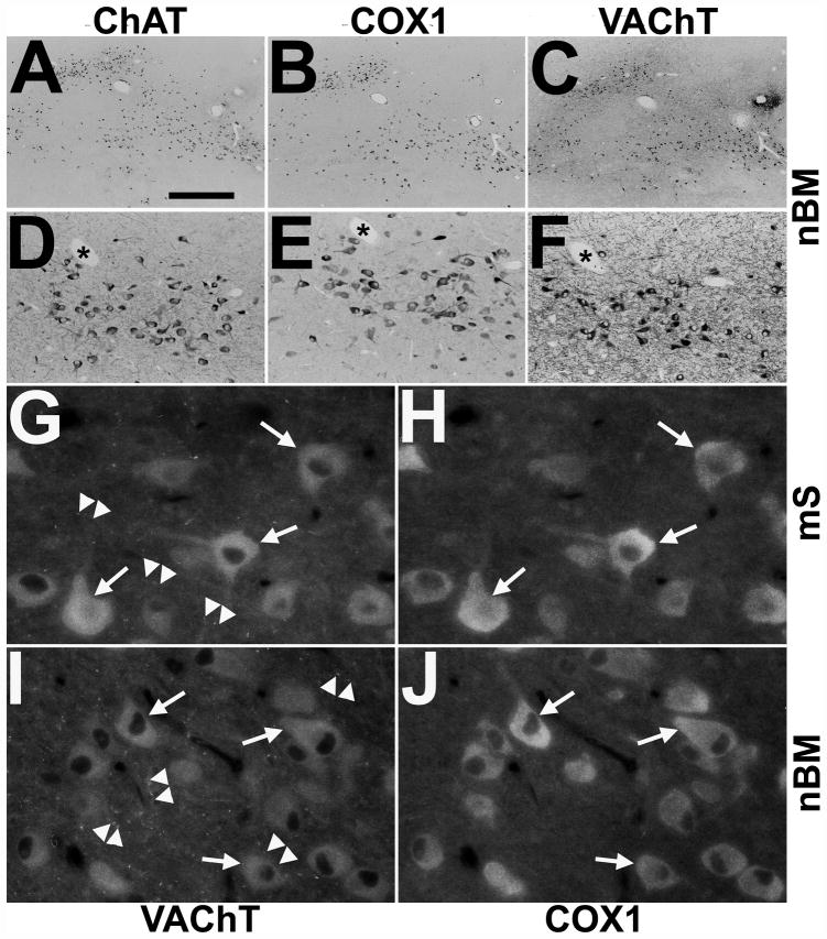
Immunodetection of cyclooxygenases 1 (COX1) in cholinergic neurons of the rhesus monkey basal forebrain. (A–F) Representative single immunohistochemistry of adjacent sections alternately stained for choline acetyltransferase (ChAT) (A, D), COX1 (B, E) and vesicular acetylcholine transporter (VAChT) (C, F) reveal COX1 in the majority of cholinergic neurons of the nucleus basalis of Meynert (nbM) of a control rhesus monkey as shown in low- (A–C) and medium-power (D–F) images. Asterisks in (D–F) mark cross-sectional profiles of the same blood vessel at different section planes. (G–J) High-power double immunofluorescence demonstrates colocalization of COX1 (H, J) with VAChT (G, I) in somata and proximal processes of cholinergic neurons (arrows) but not in VAChT-positive varicose fibers and synapses (arrowheads) in the medial septum (mS; G, H) and the nbM (I, J) of a control monkey. Scale bars: A–C, 1000 μm; D–F, 200 μm; G–J, 40 μm.

