Figure 3.
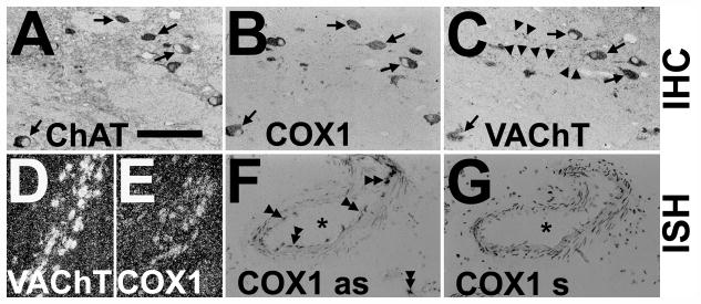
Expression of cyclooxygenase 1 (COX1) protein and COX1 mRNA in cholinergic neurons of the human basal forebrain and specificity of COX1 riboprobe. (A–C) Adjacent sections alternately immunostained (IHC) for choline acetyltransferase ChAT (A), COX1 (B) and vesicular acetylcholine transporter VAChT (C) demonstrate COX1 in cholinergic neurons (arrows) of the human nucleus basalis of Meynert. Notice that cholinergic varicose fibers are only stained for VAChT (arrowheads; C). (D, E) Representative low-power darkfield images of in situ hybridization (ISH) with [35S]-labeled riboprobes on interval sections from human nucleus basalis of Meynert reveal occurrence of COX1 mRNA (silver grains; E) in VAChT mRNA-expressing neurons (silver grains; D). (F, G) Bright-field images of riboprobe in antisense orientation against COX1 mRNA is specific and detects COX1 mRNA in endothelial cells and microglia in a human brain tissue section (double arrowheads; COX1 as; F) ISH with [35S]-labeled riboprobe in sense orientation against COX1 mRNA demonstrates no any signals except background (COX1 s, G). Asterisks show the same blood vessel in weakly hematoxylin and eosin–stained adjacent sections. Bars: A–C, 100 μm; D, E, 200 μm; F, G, 150 μm.

