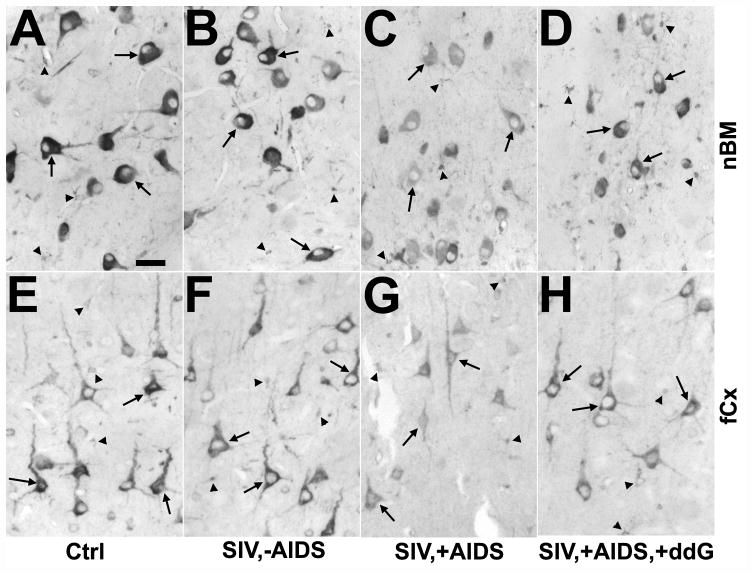
Figure 4.
Changes in neuronal cyclooxygenase 1 (COX1) immunoreactivity in the nucleus basalis of Meynert (nbM; A–D) and frontal cortex (fCx; E–H) of rhesus monkeys after SIV infection and antiretroviral treatment with 6-Cl-ddG. (A–D) The intensity of COX1 immunostaining in neurons (arrows) of the nbM is reduced in a monkey with AIDS (SIV,+AIDS) (C), as compared to a non-infected control (Ctrl) (A), and a SIV-infected monkey without AIDS (SIV,−AIDS) (B); this was not reversed by 6-Cl-ddG treatment in AIDS (SIV,+AIDS,+ddG) (D). (E–H) Reduction of neuronal COX1 immunostaining (arrows) in the fCX of an AIDS-diseased monkey (G) is reversed by 6-Cl-ddG treatment (H) to levels observed in a Ctrl (E) or a SIV,−AIDS monkey (F). There are COX1-positive microglia (arrowheads) intermingled between the neurons that demonstrate the same immunostaining intensity during disease as the neurons; the staining background in these sections is similar. Bar: 50 μm.

