Figure 6.
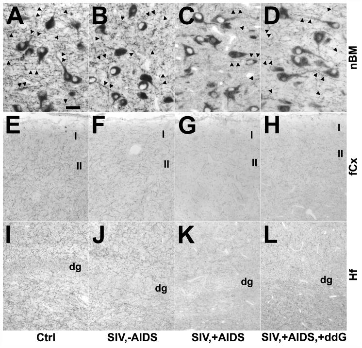
Changes in cholinergic fibers in the nucleus basalis of Meynert (nbM; A–D), frontal cortex (fCx; E–H) and hippocampal formation (Hf; I–L) of rhesus monkeys after SIV infection and antiretroviral treatment with 6-Cl-ddG as demonstrated by immunohistochemistry for the vesicular acetylcholine transporter (VAChT). (A–D) The density of VAChT-positive varicose fibers and synapses (arrows) is decreased in the nbM of a monkey with AIDS (SIV,+AIDS) (C), as compared to a non-infected control (Ctrl) (A), and a SIV-infected asymptomatic monkey (SIV,−AIDS) (B). Antiretroviral treatment with 6-Cl-ddG minimally reversed the VAChT-positive fiber loss in nbM in AIDS (SIV,−AIDS,+ddG) (D). The staining intensity of VAChT-positive neurons is not significantly altered during disease and treatment (B–D), as compared to Ctrl (A). (E–L) VAChT-positive terminals are reduced in the fCx (G) and Hf (K) of a SIV,+AIDS monkey as compared to a Ctrl monkey (E, I) and a SIV,−AIDS monkey (F, J). Treatment with 6-Cl-ddG does not prevent the reduction of VAChT-positive fibers in the fCx (H) and in the Hf (L) in late stage SIV disease. Group representative images show the upper laminae of fCx (I and II; E–H) and the area of dentate gyrus (dg; I–L). Bars: A–D, 50 μm; E–L, 200 μm.

