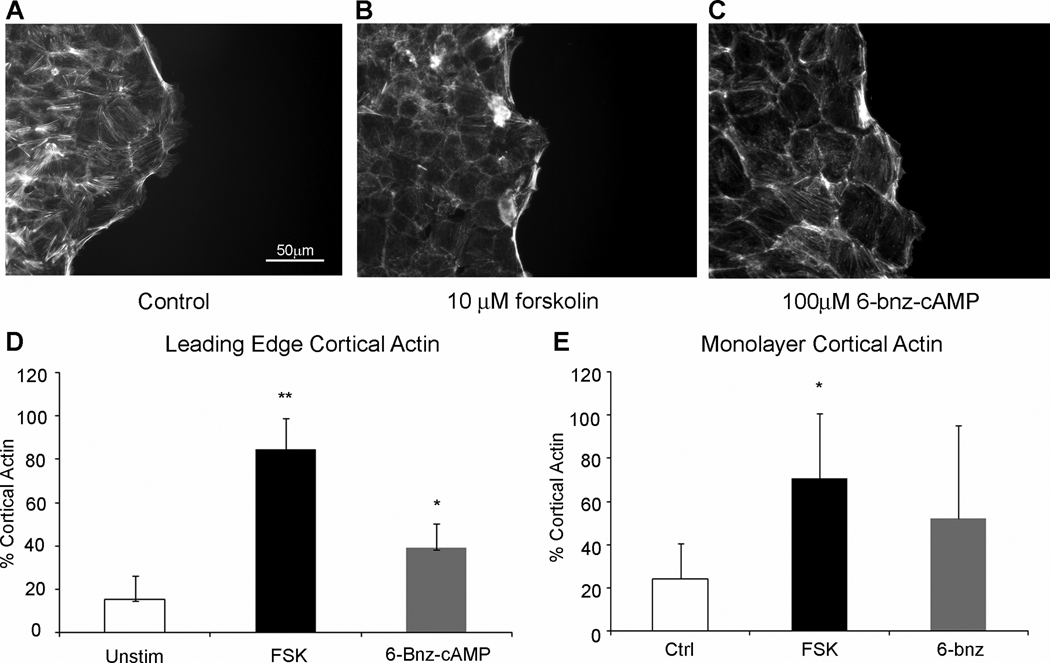
Figure 7. PKA activation decreases cell migration through altered actin regulation.
Fluorescence micrographs of wounded monolayers (A) untreated, (B) forskolin, or (C) 6-bnz-cAMP. (D) Quantification of cortical and stress fiber actin at the leading edge of the wounded epithelium. *, P≤0.05; **, P≤0.01; mean±SEM of 4 experiments. (E) Quantification of cortical and stress fiber actin at 5–10 rows back into the monolayer from the leading edge. *, P≥0.05. Images are representative of 5–7 fields of view from 4 experiments.

