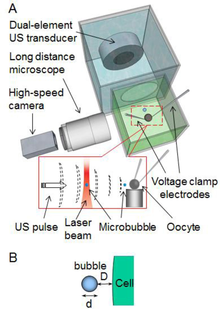
Figure 1.
Experimental setup. (A) A single oocyte was placed in ND96 solution. The femtosecond laser beam generated and trapped a single microbubble 500 µm horizontally from the oocyte. An ultrasound transducer assembly in an adjacent tank controlled and generated cavitation. Voltage-clamp measured the TMC of the cell in real time. A high speed camera was used to image the ultrasound-driven dynamic bubble activities. Inset: A microbubble generated by LIOB was trapped and then moved toward the oocyte by the 7 MHz ultrasound pulses. (B) Schematic illustration of the separation distance D between the cell membrane and a bubble (diameter of d).

