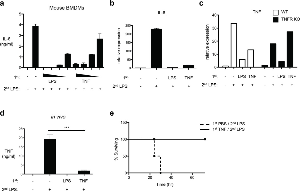
Figure 2.
TNF suppresses cytokine production in vivo and protects mice from LPS-induced lethality. (a) Mouse BMDMs were stimulated with increasing doses of LPS (1–100 ng/ml) or TNF (1–80 ng/ml) for 24 hr and then challenged with 10 ng/ml LPS. IL-6 in culture supernatants was measured by ELISA. (b) LPS or TNF-pretreated BMDMs as in (a) were stimulated with 10 ng/ml LPS for 1 hr. Real-time PCR was used to measure IL-6 mRNA levels (mean ± s.d. of triplicate determinations). (c) BMDMs from mice lacking TNFR1 and TNFR2 (TNFR KO) or genetically matched controls (WT) were stimulated with LPS (100 ng/ml) or TNF (40 ng/ml) for 24 hr and then challenged with 10 ng/ml LPS for 1 hr. TNF mRNA levels were determined by real-time PCR. Data are representative of three independent experiments. (d) Age- and sex-matched C57/BL6J mice received intraperitoneal (IP) or intravenous (IV) injection of LPS (100 µg) or of TNF (2 µg), respectively. After 24 hr, secondary LPS challenge (200 µg) was given by IP injection and after 90 min serum TNF was determined by ELISA (n=4 mice per group). P value was calculated by unpaired Student’s t-test. ***, P = 0.0004. Similar results were obtained in an additional 2 experiments using different TNF dosing regimens. (e) Mice were injected intravenously with TNF (2 µg), followed by injection of 500 µg of LPS after 24 hr. Survival rates were scored every 6 h for 4 days (n=4 mice per group). Similar results were obtained in an additional 3 experiments using different TNF pretreatment doses.

