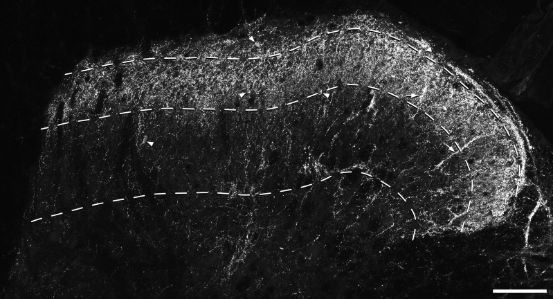
Figure 2.
NPY-immunostaining in the dorsal horn. A confocal image from a transverse section through the dorsal horn immunostained to reveal NPY. The dashed lines represent the ventral borders of laminae I, II, and III. There is a dense plexus of immunoreactive axons that occupies laminae I and II, with some axonal staining in deeper laminae. Dorsoventrally orientated bundles of axons can often be seen and two of these are marked with arrows. Scattered immunoreactive cell bodies are present throughout laminae I–III and some of these are indicated with arrowheads. Note that many of those in laminae I–II are hidden by the axonal plexus. The image is a projection of 24 optical sections at 1-μm z-spacing. Scale bar = 100 μm.

