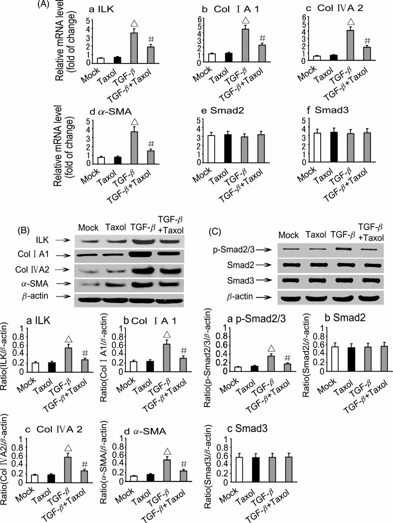
Figure 4.
Effect of Taxol on ILK, Collagen, phosphorylated Smad2/3 (p-Smad), Smad2, Smad3 and α-SMA expression in NRK52E cells. (A) Real-time PCR analyses showed that ILK, Collagen and α-SMA were significantly higher in the TGF-β1 group compared to the Mock group (p < 0.05, n = 6). Treatment with Taxol caused a significant decrease in their expression in TGF-β1-treated NRK52E cells (p < 0.05, n = 6). Smad2 and Smad3 mRNA expression were unaltered in the Taxol and TGF-β1 + Taxol groups. (B, C) Western blot analyses showed that ILK, Collagen, p-Smad2/3 and α-SMA, but not total Smad2 or Smad3, expressions were significantly higher in the TGF-β1 group compared to the Mock group (p < 0.05, n = 6), while treatment with Taxol reduced their expression (p < 0.05, n = 6). Δp < 0.05 versus Mock or Taxol groups (n = 6); #p < 0.05 versus TGF-β1 group

