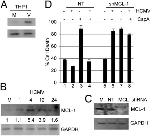
Fig. 2.
HCMV up-regulation of MCL-1 correlates with protection from cell death. (A and B) MCL-1 and GAPDH protein expression was analyzed in mock- (M) and HCMV (V)-infected cells 4 hpi of THP1 cells (A) and 0–24 hpi were analyzed in CD34+ cells infected with HCMV (B). Densitometry shows the ratio change from mock. (C) Western blot for MCL-1 and GAPDH expression was performed on THP1 cells 24 h after transduction with mock (M) or a nontargeting (NT) or shMCL-1 expressing (MCL) lentiviral vector. (D) THP1 cells were first transduced with either nontargeting (bars 1–4) or shMCL-1–expressing (bars 5–8) lentiviral vectors for 24 h and then mock- or HCMV-infected. At 2 hpi, they were incubated with DMSO or cisplatin A for 4 h. Following rescue in fresh media, cell viability was assayed by trypan blue staining 18 h after cisplatin A treatment.

