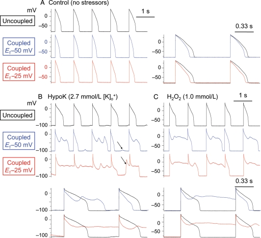
Figure 2.
Promotion of EADs by myofibroblast–myocyte coupling. (A) Coupling a patch-clamped myocyte superfused with normal Tyrode's solution to a virtual fibroblast did not induce EADs at PCL 1 s. Superimposed traces at a faster time scale to the right illustrate that coupling lowers the AP plateau and shortens ADP. (B and C) In myocytes exposed to hypokalaemia or H2O2 to induce bradycardia-dependent EADs at PCL 6 s (not shown), pacing at PCL 1 s suppressed EADs completely (top traces). However, coupling the myocyte to a virtual fibroblast (Cf 6.3 pF, Gj 3.0 nS) caused EADs to reappear, which were more prominent when Ef was −25 mV (bottom traces) than −50 mV (middle traces). In addition to EADs, DADs (some triggering APs) were also frequently observed (arrows). Lower panels show superimposed traces of control vs. coupled for the Ef −50 and −25 mV cases, respectively, at a faster time scale. Note that coupling lowers the early AP plateau voltage before EAD onset.

