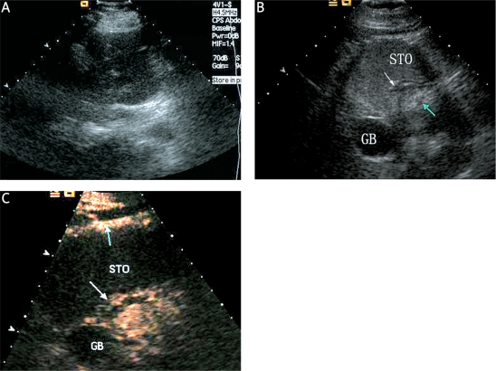
Figure 2.
A case of EGC without LNM confirmed pathologically after operation. A: The early gastric cancer and LNM could not be detected with conventional transabdominal US. B: The early gastric cancer (white arrow) and LNM (green arrow) can be clearly demonstrated in the oral contrast-enhanced ultrasonography image. C: Compared with the adjacent normal gastric wall (white arrow), unmarked hyperenhancement of the EGC (green arrow) in DCUS image was shown during the early arterialphase
STO – stomach, GB – gallbladder

