Fig. 4.
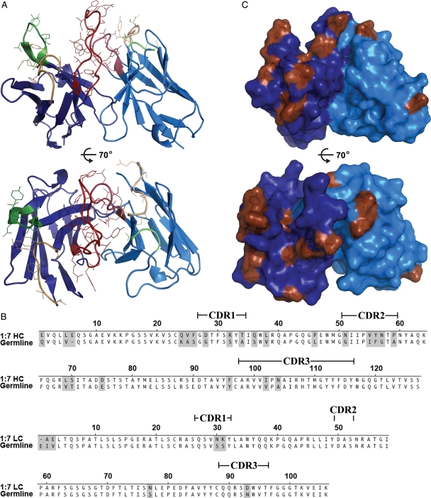
Crystal structure of scFv 1:7. The crystal structure of scFv 1:7 (A) viewed from the side (upper panel) and from the top (lower panel). Framework regions are colored in dark blue (HC) or light blue (LC) and the CDRs according to the immunogenetics (IMGT) nomenclature are colored in yellow (CDR1), green (CDR2) and red (CDR3). The side chains of the residues in the CDRs are shown as lines. (B) The 1:7 amino acid sequence was aligned with the closest homologous germline sequence suggested by IMGT V-QUEST and junction analysis (Lefranc et al., 2009). Positions of somatic mutations during antibody maturation are shaded, bars indicate the positions of the CDRs according to the IMGT nomenclature. (C) Mapping of the somatic mutations (brown) on the molecular surface of scFv 1:7 from the side (upper panel) and from the top (lower panel).

