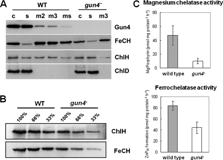
FIGURE 3.
Localization, quantification, and activities of both chelatases. A, subcellular localization of Gun4, ferrochelatase (FeCH), and the magnesium chelatase ChlH and ChlD subunits. Membranes have been washed 2 (m2) or 3 (m3) times in thylakoid buffer followed by an additional wash with 2 m NaCl in thylakoid buffer (ms). Membrane fractions (m), crude (c), and soluble extracts (s) of wild-type and gun4 mutant cells were separated by SDS-PAGE and transferred to nitrocellulose membranes. The amount of Chl corresponding to 150 μl of cells at A730 = 1 was loaded per each lane. Polyclonal antibodies used for the immunoblots are shown on the right. B, accumulation of the ChlH subunit of magnesium chelatase and ferrochelatase in the crude cell extract were determined by immunodetection. Numbers indicate relative protein loading based on the amount of cells. C, activities of magnesium and ferrochelatase measured in wild-type and gun4 mutant cells.

