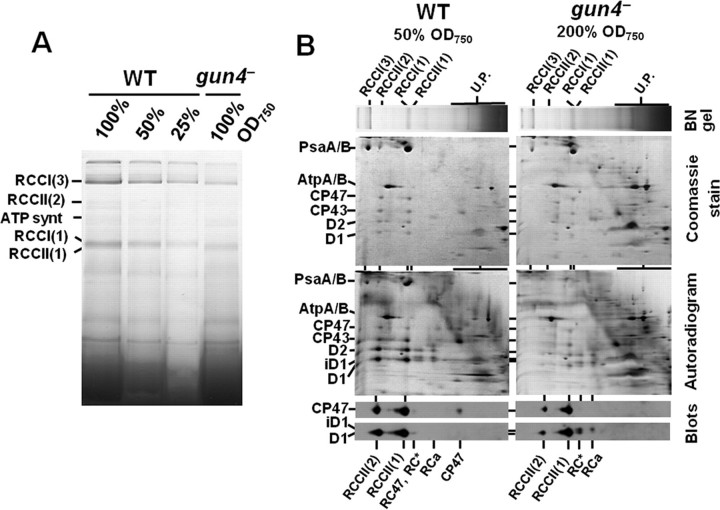
FIGURE 5.
Analysis of membrane protein complexes and their assembly in wild-type and gun4 mutant cells. A, membrane protein complexes from wild-type and gun4 mutant labeled in vivo with 35[S]Met/Cys were separated in 4–16% BN gel as described under “Experimental Procedures.” Amounts of Chl corresponding to designated amounts of cells were loaded per each lane. B, two-dimensional analysis of membrane protein complexes separated in A. Equal amounts of Chl corresponding to the half (50% A750) and double (200% A750) amounts of wild-type and gun4– cells were loaded, respectively. Lanes from the BN-gel were excised and placed on top of the 12–20% denaturating gel. The two-dimensional gels were either stained by Coomassie Blue (Coomassie gels), dried, and exposed to phosphorimaging plates (Autoradiograms) or electroblotted onto a polyvinylidene difluoride membrane that was immunodecorated using antibodies raised against D1 and CP47 proteins (blots). Designation of complexes: RCCI(3) and RCCI(1), trimeric and monomeric PSI complexes, respectively; RCCII(2) and RCCII(1), dimeric and monomeric PSII core complexes, respectively; RC47, the monomeric PSII core complex lacking CP43; RC* and RCa, reaction center complexes accumulating in PSII mutants lacking CP47 (24); U.P., unassembled proteins; iD1 designates D1 processing intermediate.

