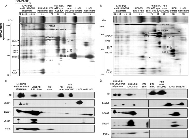
FIGURE 1.
Two-dimensional analysis of protein complexes composition of mesophyll (A and C) and bundle sheath (B and D) thylakoids of maize. Thylakoid protein samples (50 μg of Chl) were solubilized in the presence of 1 and 2% DDM for MS and BS thylakoids, respectively, and loaded onto BN gels (4–10% acrylamide, 4–15% sucrose). For second dimension, lanes were cut out of the BN gels and loaded horizontally onto denaturing SDS-PAGE gels (15% acrylamide, 6 m urea). Protein bands were visualized by Coomassie staining (A and B) or immunodetected (C and D) using antibodies against selected subunits of PSI (PSI L, Lhca1, and Lhca4) and PSII (D2 and Lhcb1). Protein bands resolved on BN-PAGE are numbered above the BN gel fragments. w/o CP43, without CP43. Molecular masses of protein standards (in kDa) are indicated on the left. The black line in Fig. 1D indicates the joining point of the two pieces of the membrane. Note that the immunodetection analysis is not quantitative, since proteins were blotted onto separate membranes.

