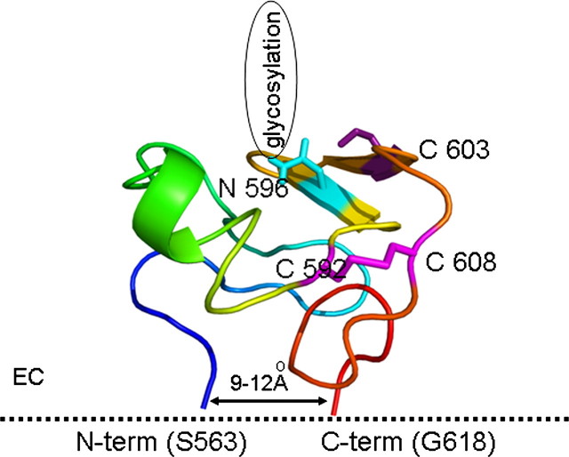
FIGURE 8.
Model of the third extracellular loop of ABCG2 (amino acids 563–618) obtained by ab initio folding employing discrete molecular dynamics. The structure is colored blue to red from the N to the C terminus. Cysteines involved in intramolecular and intermolecular interactions are represented with sticks and are colored magenta. The glycosylation site (Asn-596) is represented by a blue stick.

