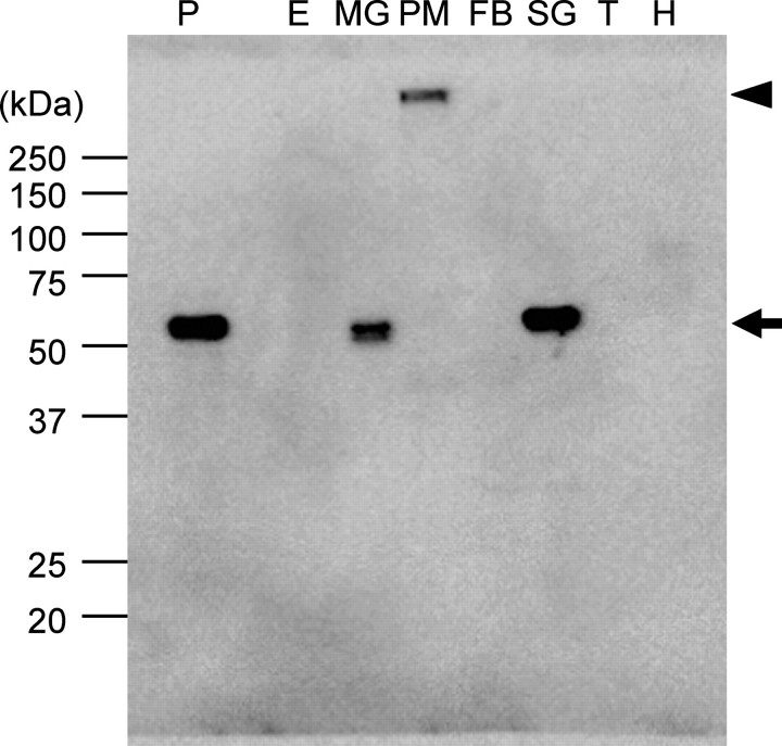
FIGURE 6.
Immunoblot analysis of BmSUC1 protein. Immunoblot analysis using anti-BmSUC1 antiserum was performed. Protein samples were isolated from the epidermis (E), midgut (MG), soluble fraction (supernatant of the homogenate) of peritrophic membrane (PM), fat bodies (FB), silk gland (SG), trachea (T), and hemolymph (H) from the 3-day-old fifth instar larvae. 2 μg of protein was loaded in each lane. As a positive control, the medium from cells infected with AcMNPV that expressed BmSUC1 protein was also loaded (P). The size and position of the protein standards are indicated on the left. BmSUC1 protein signals are shown by an arrow. An arrowhead shows the signal from putative BmSUC1 protein from the peritrophic membrane, which did not enter the running gel.

