FIGURE 7.

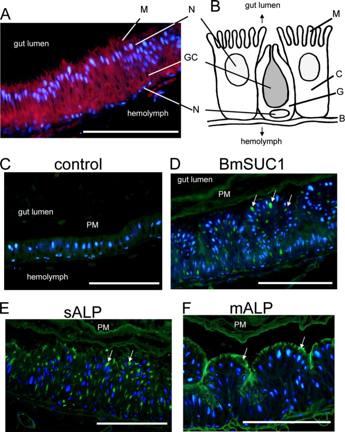
Immunohistochemical analysis of BmSUC1 protein in the midgut. A, midgut cells stained with DAPI (blue) and AlexaFluor594-conjugated phalloidin (red). B, schematic representation of midgut cells of B. mori. C, columnar cell; G, goblet cell; GC, goblet cell cavity; N, nucleus; B, basal lamina; M, microvilli. C, control experiments using preimmune serum. D, immunofluorescence visualization of BmSUC1. Sections were incubated with anti-BmSUC1 antibody followed by the secondary antibody labeled with AlexaFluor488 (green) and counterstained with DAPI. Strong signals were detected in the goblet cell cavities. E and F, control experiments using antisera against sALP (green) and mALP (green), respectively. Note that localization of BmSUC1 is closely similar to that of sALP, which was shown to localize within goblet cell cavities. Arrows, localization of the protein; bar, 200 μm.
