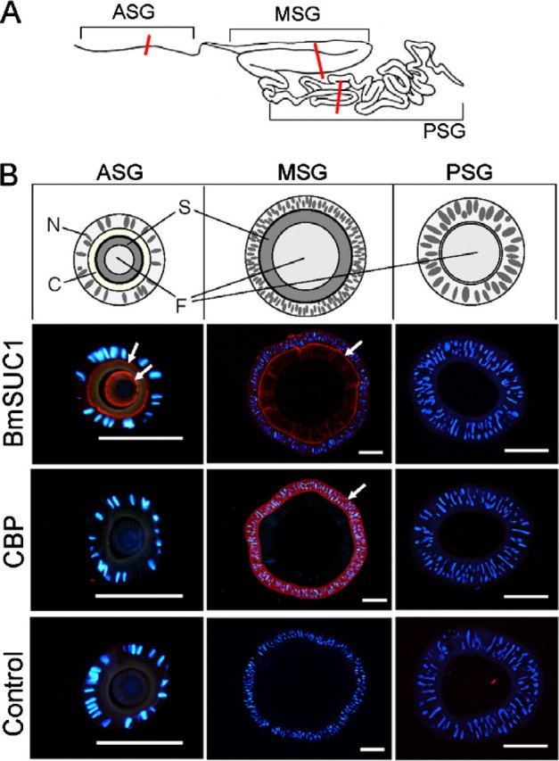
FIGURE 8.

Immunohistochemical analysis of BmSUC1 protein in the silk gland. A, schematic representation of the silk gland of the silkworm. Sections were obtained from the middle part of the anterior silk gland (ASG), MSG, and posterior silk gland (PSG), as indicated by red lines. B, immunofluorescence visualization of BmSUC1 and CBP. Slides were incubated with anti-BmSUC1 or anti-CBP antiserum followed by the secondary antibody labeled with AlexaFluor546 (red) and counterstained with DAPI (blue). Control experiments were also preformed using preimmune serum. Arrows, localization of the protein; bar, 200 μm. Schematic representations of sections are also shown (top). N, nucleus; C, cuticle layer; S, sericin layer; F, fibroin layer.
