
FIGURE 1.
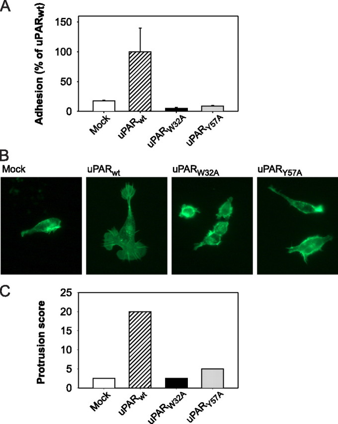
Vitronectin adhesion and cytoskeletal rearrangements in HEK cells expressing wild-type and mutated uPAR. A, adhesion of uPAR transfected cells on a reconstituted vitronectin matrix. Mock-transfected (Mock) cells or cells expressing uPARWT or uPAR mutant proteins were seeded in vitronectin-coated culture wells in the presence of EDTA and allowed to adhere during a 1-h incubation period at 37 °C. After washing, adherent cells were quantified by thiazolyl blue tetrazolium bromide assay. Each column represents the mean of a triple determination. The standard deviations are indicated. B, morphological changes and cytoskeletal rearrangements. Mock-transfected cells or cells expressing uPARWT or uPAR mutant proteins were cultured for 5 days on vitronectin-coated coverslips. The cells were then fixed and permeabilized followed by FITC-phalloidin staining and examination by fluorescence microscopy. Note the exclusive appearance of lamellipodia and cytoskeletal extensions in the uPARWT-transfected cells. C, quantification of fields with lamellipodia-positive cells. Cells were cultured, stained, and examined by fluorescence microscopy as in B. Each cell type was assigned an arbitrary designation, after which five randomly selected microscope fields for each cell type were scored blindly by four investigators for lamellipodia-positive cells (see “Experimental Procedures”). The cumulative score is represented for each sample.