
FIGURE 10.
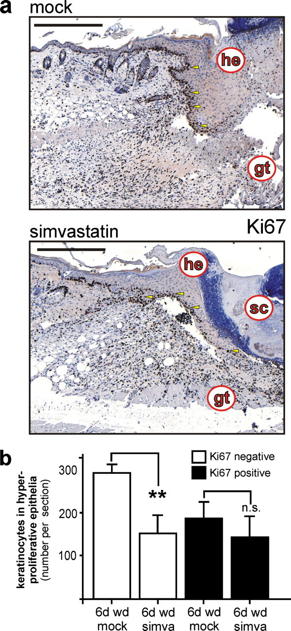
Proliferation of wound keratinocytes in statin-treated mice. a, analysis of keratinocyte proliferation using a rat monoclonal antibody directed against the proliferative antigen marker Ki67 in 6-day wound tissue of untreated (mock) and simvastatin-treated (simva) mice as indicated. b, quantitative analysis of total and Ki67-expressing wound keratinocyte cell numbers from stained wounds. **, p < 0.01; n.s., not significant (unpaired Student's t test) as indicated by brackets. Bars indicate the mean ± S.D. obtained from wounds (n = 6) isolated from three individual animals (n = 3). Sections were stained with the avidin-biotin-peroxidase complex system using 3,3-diaminobenzidine-tetrahydrochloride as a chromogenic substrate, and nuclei were counterstained with hematoxylin. gt, granulation tissue; he, hyperproliferative epithelium; sc, scab. Bars, 100 μm.