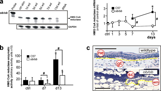
FIGURE 11.
Impaired regulation of HMGR in diabetes-disturbed skin repair. a, RNase protection assay for HMGR mRNA expression in skin wounds of diabetic ob/ob mice. The time after injury is indicated at the top of each lane. ctrl skin refers to back skin biopsies of non-wounded mice. 1000 cpm of the hybridization Probe were used as a size marker. Hybridization against GAPDH was used as a loading control. A quantification of HMGR mRNA (PhosphorImager PSL counts per 15 μg of total wound RNA) is shown in the right panel. *, p < 0.05 (ANOVA, Dunnett's method) compared with control skin. #, p < 0.05 (unpaired Student's t test) compared with wild-type (C57) mice. Bars indicate the mean ± S.D. obtained from wounds (n = 48) isolated from animals (n = 12) from three independent animal experiments. b, HMGR activity assays of wound tissue from wild type (C57) and diabetic ob/ob mice (ob/ob) as assessed using 3-methyl [3-14C]glutaryl coenzyme A as substrate. * and §, p < 0.05 (ANOVA, Dunnett's method) compared with the respective control. #, p < 0.05 (unpaired Student's t test) as indicated by brackets. Bars indicate the mean ± S.D. obtained from wounds (n = 24) isolated from animals (n = 12) from three independent animal experiments. c, hematoxylin-counterstained representative sections from 13-day wound tissue of wild-type and diabetic ob/ob mice as indicated. Epithelia are highlighted with the yellow line. gt, granulation tissue; ne, neo-epithelium; sc, scab; wm, wound margin. Bars, 50 μm.

