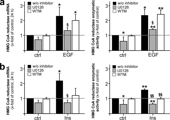
FIGURE 5.
Dependence of EGF- and insulin-mediated HMGR expression on MAPK and PI3K activation. Quiescent human HaCaT keratinocytes were stimulated with EGF (a) or insulin (b) for 24 h in the absence or presence of U0126 (10 μm) or wortmannin (WTM, 200 nm) as indicated and subsequently analyzed for HMGR mRNA expression by RNase protection assay (left panels) and activity using 3-methyl [3-14C]glutaryl coenzyme A as substrate (right panels). **, p < 0.01; *, p < 0.05 (unpaired Student's t test) as compared with the respective control. §§, p < 0.01; §, p < 0.05 (unpaired Student's t test) as compared with stimulated cells w/o inhibitor. Bars indicate the mean ± S.D. obtained from three (n = 3) independent cell culture experiments.

