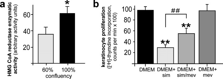
FIGURE 8.
HMGR activity in the control of keratinocyte proliferation. a, HaCaT keratinocytes were grown to 60% or 100% confluency, respectively. Cells were subsequently lysed and HMGR activity was assessed using 3-methyl [3-14C]glutaryl coenzyme A as substrate. *, p < 0.05 (unpaired Student's t test) as compared with control. Bars indicate the mean ± S.D. obtained from three (n = 3) independent cell culture experiments. b, HaCaT keratinocytes were grown exponentially in Dulbecco's modified Eagle's medium (DMEM) and treated with simvastatin (10 μm), mevalonate (1 mm), or a combination of both as indicated for 24 h. Non-treated cells served as control (DMEM). Proliferation rates were assessed by analysis of [3H]thymidine incorporation. **, p < 0.01 (unpaired Student's t test) as compared with control (DMEM). ##, p < 0.01 as indicated by the bracket. Bars indicate the mean ± S.D. obtained from three (n = 3) independent cell culture experiments.

