
FIGURE 2.
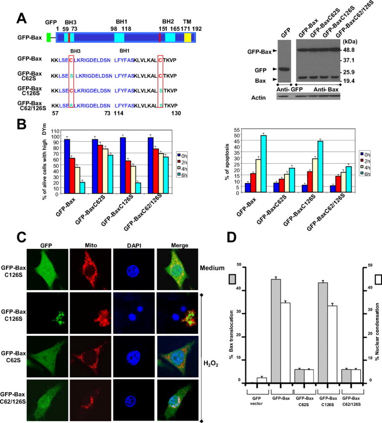
Cysteine 62 to serine substitution prevents Bax translocation and apoptosis. A, left, schematic overview of GFP-Bax and GFP-Bax mutant constructs. Right, expression levels of GFP-Bax and its mutants in SW480 cells. Cell lysates were subjected to SDS-PAGE, blotted, and probed with the indicated antibodies. B, cells were exposed to H2O2 (25 μm) for indicated times. Left, the percentage of living cells with high mitochondrial membrane potential was determined by 3,3′-dihexyloxacarbocyanine iodide and propidium iodide staining, followed by flow cytometry. Right, apoptotic cells were identified as Annexin V+ cells. C, confocal microscopy analysis of translocation of GFP-Bax mutants to mitochondria. Cells were cultured in the presence or absence of 25 μm H2O2 for 6 h and then subjected to mitochondrial fluorescence staining with 50 nm MitoTracker. Nuclei were counterstained with DAPI. D, graphs showing results of quantitative analyses. Apoptotic cells were characterized by nuclear condensation. At least 200 cells were counted in each experiment (n = 3, mean ± S.D.).