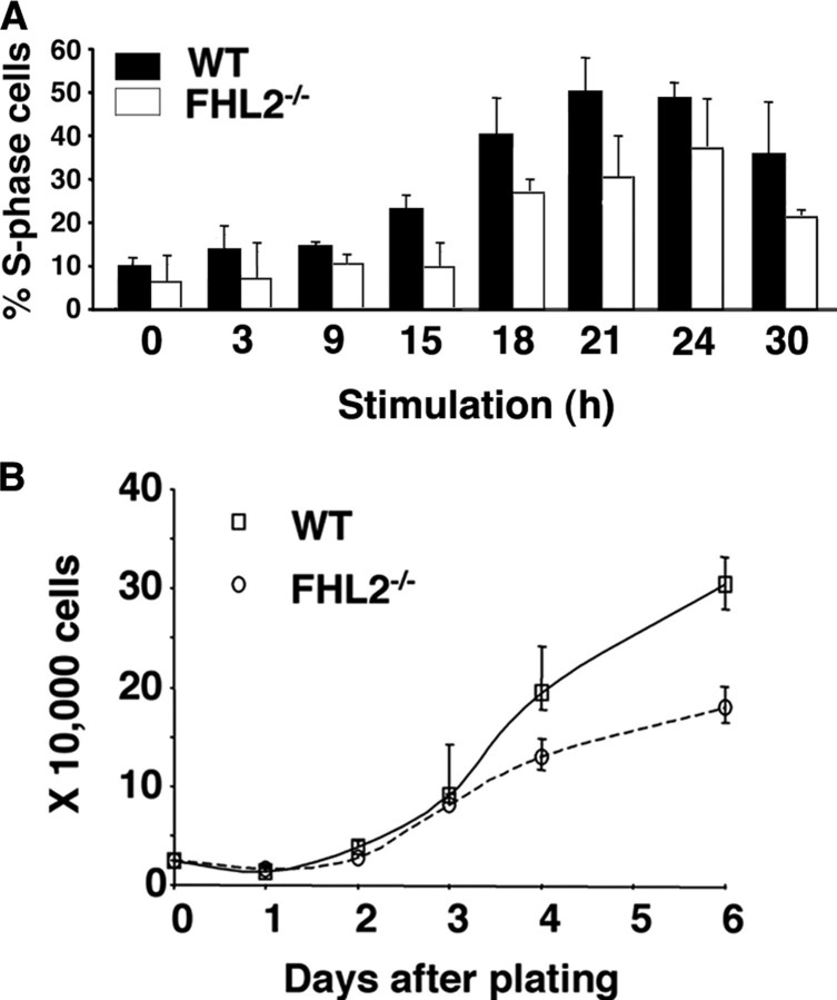
FIGURE 3.
Cell proliferation in immortalized WT and FHL2-/- MEFs. A, kinetics of S phase entry in immortalized MEFs. After serum stimulation, cells were pulse-labeled with BrdUrd and harvested at the indicated time of stimulation. The percentage of cells in S phase at each time point was calculated and plotted based on flow cytometry results of three independent experiments. B, decreased cell proliferation in immortalized FHL2-/- cells. Equal numbers of spontaneously immortalized WT and FHL2-/- MEFs were plated in triplicate. Cell numbers were determined during 6 days. The data presented are the mean value ± S.D. obtained from three independent experiments. Data shown are representative of growth curves of three independent clones for each genotype.

