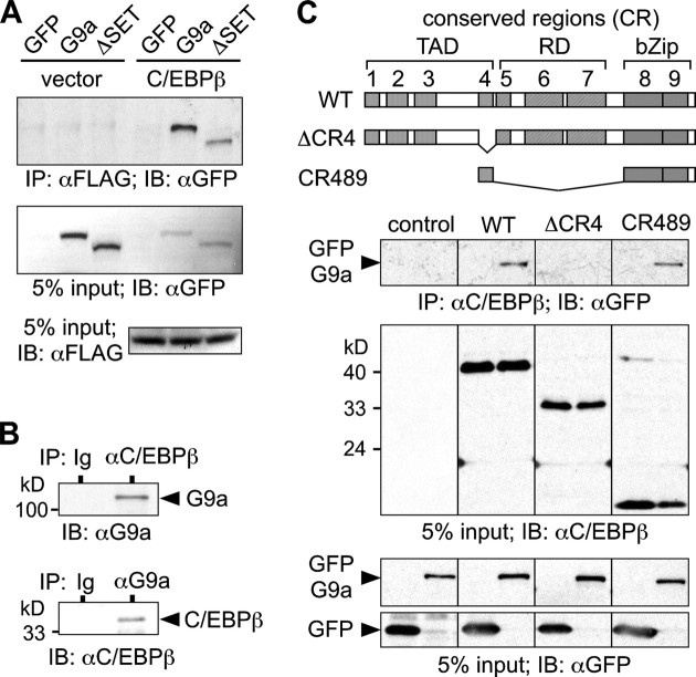
FIGURE 1.
C/EBPβ and G9a interact in eukaryotic cells. A, empty vector or pcDNA3-C/EBPβ LAP*-FLAG was transfected together with pCMV-GFP, pCMV-GFP-G9a, and pCMV-GFP-G9aΔSET, as indicated, into HEK-293 cells and immunoprecipitated (IP) using anti-FLAG affinity matrix. Bound proteins were analyzed by immunoblotting (IB) using anti-GFP antibodies. Protein input was monitored with anti-FLAG or anti-GFP antibodies. B, Jurkat cells were lysed and immunoprecipitated with anti-C/EBPβ, anti-G9a, or Ig (control) antibodies as indicated. Bound proteins were analyzed by immunoblotting. C, structure of C/EBPβ, indicating TAD, regulatory domain (RD), and CR, is shown. pcDM8-C/EBPβ LAP*, pcDM8-C/EBPβ ΔCR4, pcDM8-C/EBPβ CR489, and pCMV-GFP-G9a constructs were transfected into HEK-293 cells and immunoprecipitated using anti-C/EBPβ antibody and protein A-Sepharose and revealed by immunoblotting, as indicated. WT, wild type; bZIP, basic leucine zipper.

