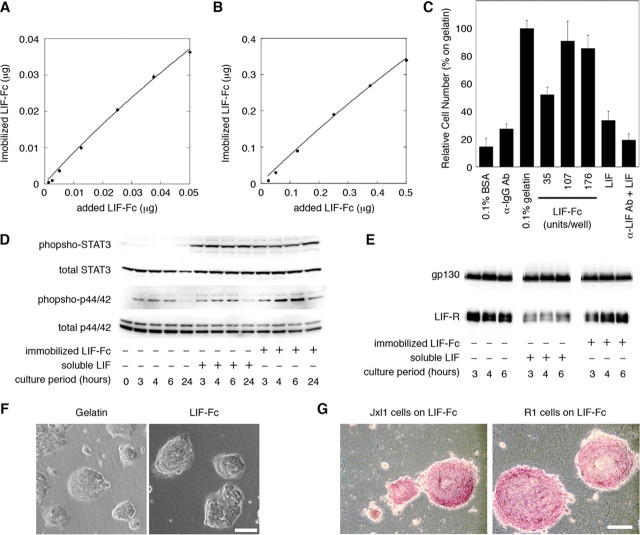
FIGURE 3.
The amount of immobilized LIF-Fc was measured at a low concentration (A) and a high concentration (B) of LIF-Fc. The data indicate means ± S.E. of five separate experiments. C, adhesion of mouse ES cells onto the surface of 96-well plate coated with 0.1% BSA, anti-mouse IgG antibody, 0.1% gelatin, LIF-Fc, recombinant LIF (10,000 units/ml), or anti-LIF antibody followed by recombinant LIF (10,000 units/ml). After 3 h of incubation, ES cells adhered to a LIF-Fc-coated surface with equivalent efficiency as to a 0.1% gelatin-coated surface. The data indicate means ± S.D. of three separate experiments. D, activation of STAT3 and MAPK by immobilized LIF-Fc was analyzed by Western blotting. The phosphorylation of MAPK was sustained as long as 24 h after culturing. E, internalization of the LIF receptor and gp130 was analyzed by Western blotting. The amount of LIF receptor did not change on the LIF-Fc immobilized surface. F, morphological observation of R1 cells cultured on various substrates. G, ALP activity of ES cells cultured for 4 days in the 24-well plate immobilized with LIF-Fc. ES cells formed aggregated colonies on LIF-Fc-coated surface, and they maintained ALP activity without LIF supplementation. Scale bar indicates 100 μm.

