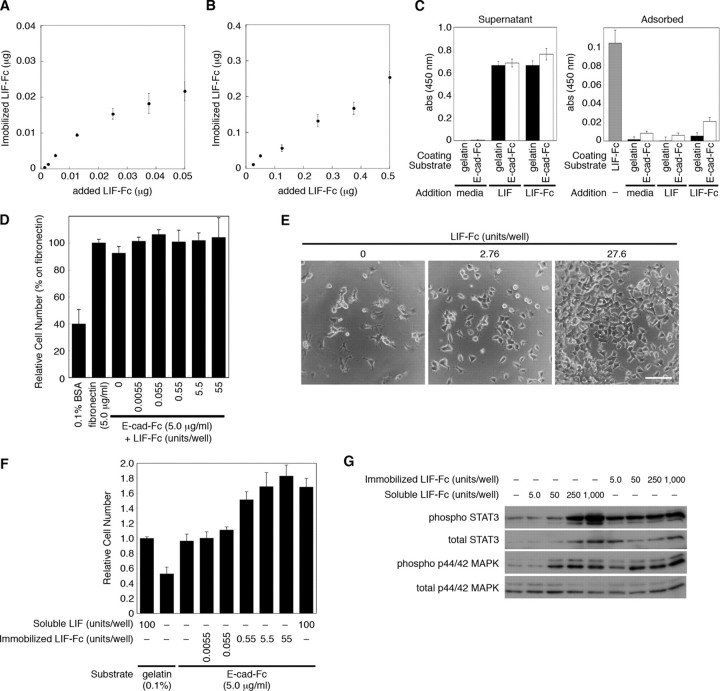
FIGURE 4.
The amount of immobilized LIF-Fc was measured at a low concentration (A) and a high concentration (B) of LIF-Fc co-immobilized with 0.25 μg of E-cad-Fc. The data indicate means ± S.E. of five separate experiments. C, amount of adsorbed LIF-Fc protein onto gelatinized or E-cad-Fc-coated surface was analyzed by ELISA. LIF or LIF-Fc was diluted with the culture media for ES cells at 1,000 units/ml and incubated on the gelatinized or E-cad-Fc-coated surface for 3 days. The amount of adsorbed or noninteracted proteins (Supernatant) was analyzed by ELISA. The surface coated with LIF-Fc (1,000 units/ml) was used as a control to compare the amount of immobilized LIF-Fc. The data represent means ± S.E. of 12 separate experiments. D, adhesion of mouse ES cells onto the surface of 96-well plate coated with 0.1% BSA, 5.0 μg/ml fibronectin, or co-immobilization of 5.0 μg/ml E-cad-Fc and various concentrations of LIF-Fc. After 3 h of incubation ES cells adhered to the co-immobilized surface with equivalent efficiency as to the surface coated with E-cad-Fc alone. The data indicate means ± S.D. of three separate experiments. E and F, ES cells show higher proliferation ability on the co-immobilized surface with E-cad-Fc and LIF-Fc, depending on the dose of LIF-Fc. E, morphological observation of ES cells cultured in the 24-well plate; the surfaces were co-immobilized with E-cad-Fc and various concentrations of LIF-Fc. ES cells scattered each other, and they proliferated dose-dependently. Scale bar indicates 100 μm. F, proliferative activity of ES cells on an indicated surface was evaluated by staining with Alamar Blue reagent. The data indicate means ± S.D. of three separate experiments. G, after culturing for 3 days in a 12-well plate, the activation of STAT3 and MAPK by immobilized LIF-Fc was analyzed by Western blotting.

