
FIGURE 1.
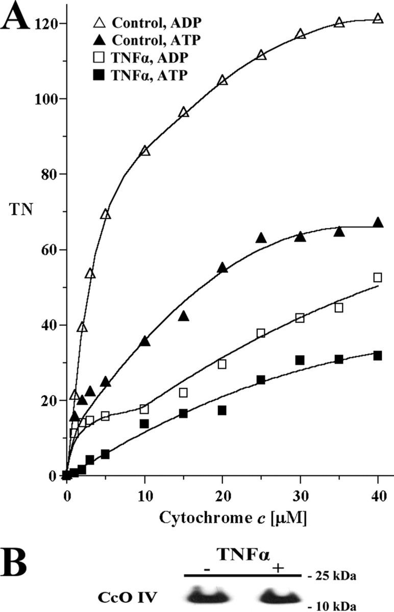
TNFα leads to CcO inhibition in cow liver tissue. A, cow liver tissue was partially homogenized leaving most cells intact and incubated in the presence (squares) or absence (triangles) of 20 ng/ml TNFα for 5 min at 20 °C. CcO activity was measured in the presence of 5 mm ATP (closed symbols, inhibited) or ADP (open symbols, stimulated) by the addition of increasing amounts of cytochrome c. Specific activity is defined as consumed O2 (μmol)/(min·total protein (mg)). Shown are representative results of three independent experiments. TN, turnover number. B, Western analysis using an antibody against CcO subunit IV (isoform 1) indicates no changes of CcO amount after short term TNFα treatment. The mitochondria were isolated with or without TNFα treatment (see above). Thirty μg of total mitochondrial protein was loaded, and CcO subunit IV was detected with a monoclonal antibody.