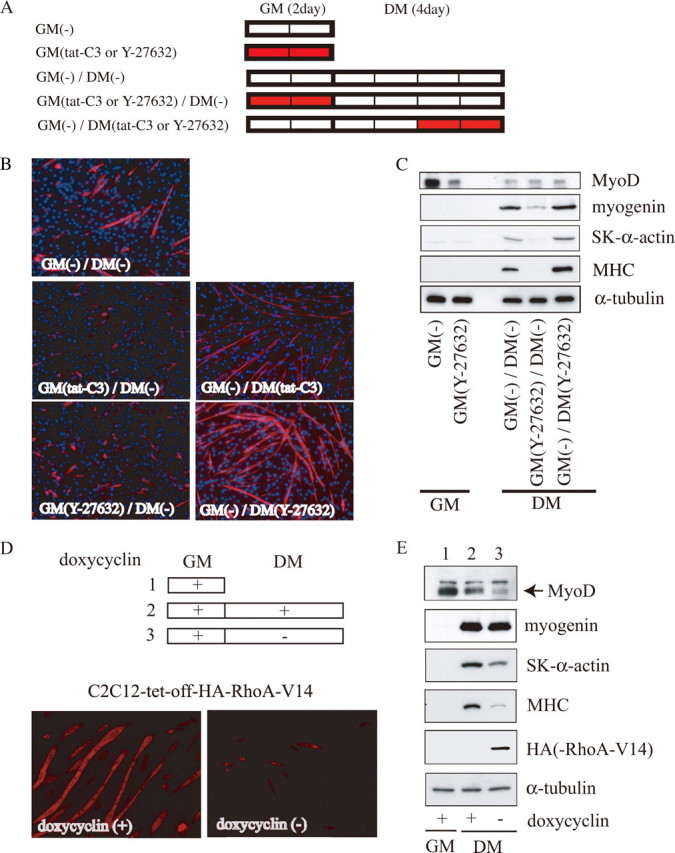
FIGURE 1.

Biphasic roles of RhoA/ROCK signaling in myogenic differentiation. A, experimental procedures are presented schematically. C2C12 myoblasts were cultured in GM for 2 days and then in DM for 4 days, to induce myogenic differentiation. The red bars show the duration of treatment with the indicated inhibitors (5 μg/ml tat-C3 or 25 μm Y-27632). B, cells (4-day cultures in DM) were stained using an anti-MHC antibody (red) and Hoechst 33258 (blue). Magnification is ×100. C, expression of myogenic differentiation markers is shown. The whole-cell extract from C2C12 cells cultured under the indicated conditions was subjected to immunoblotting using the indicated antibodies. The protein content in each sample was normalized to the amount of α-tubulin protein. Representative results from at least three experiments are shown. D, effect of the forced activation of RhoA on differentiating myocytes. The experimental procedures are presented schematically at the top. Stable C2C12 transfectants (C2C12-tet-off-HA-RhoA-V14) were cultured in GM containing 1 μg/ml doxycycline for 3 days (lane 1) and then in DM for 5 days with (lane 2) or without (lane 3) 1 μg/ml doxycycline. The cells (5-day cultures in DM) were stained using the anti-MHC antibody (bottom). Magnification is ×100. E, expression of myogenic differentiation markers is shown. The whole-cell extract from C2C12 cells cultured under the indicated conditions was subjected to immunoblotting using the indicated antibodies, as described in the legend for Fig. 1C. Lanes 1-3 correspond to those in D.
