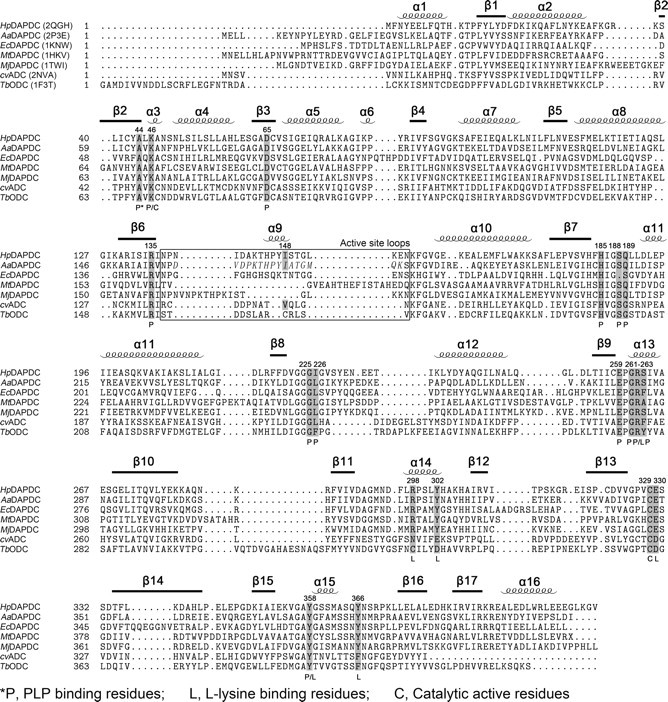
FIGURE 4.

Structure-based sequence alignment of DAPDCs, TbODC and cvADC. PDB codes are included in parentheses beside the protein names. Secondary structures of HpDAPDC are indicated above the alignment. Functional residues are shaded and labeled according to the footnote below the alignment. The italic letters in the boxed region indicate the crystallographically unobservable residues of AaDAPDC. The alignment was produced by STRAP (43) using the combinatorial extension (CE) method (44).
